At The Valley Hospital Fertility Center, compassionate care meets leading-edge expertise. With decades of experience, our dedicated team is committed to guiding you through your fertility journey with personalized care that recognizes your unique needs. At Valley, you will benefit from our innovative fertility treatments and advanced technology, including our state-of-the-art embryology lab, combined with the warmth and support of a team that truly cares about your success.
Fertility Treatment Options
Valley’s fertility team offers advanced treatment options based on your individual needs and desires. Treatment options include:
Fertility Assessment
Our center approaches fertility assessment comprehensively, by looking at the full picture of your physical and mental health. At Valley, we offer fertility assessment and infertility tests for men and women to identify any potential issues.
Infertility Treatment
After fertility assessment, our team will work with you to find the best option, based on your results and your wishes. At Valley, we offer various infertility treatment options including ovulation induction, intrauterine insemination (IUI), in vitro fertilization (IVF), preimplantation genetic testing, and more.
Third-Party Reproduction
For individuals and couples needing assistance through gestational surrogacy, donor egg services, donor sperm services, or other third-party reproduction options, Valley's Fertility Center offers the support, guidance, and services you need.
Fertility Preservation
Fertility preservation allows patients who may not be ready or able to start their family a way to preserve their fertility for the future. Due to the impact that aging has on a woman’s egg quantity and quality, freezing eggs allows patients to preserve their fertility at younger ages to maximize their chance of having a biological child in the future.
Oncofertility
Cancer treatment involving chemotherapy, radiation, and surgery can compromise a patient’s reproductive potential. Fertility preservation with egg or embryo freezing can be used before these treatments and give patients peace of mind for growing their future family.
The Valley Hospital Fertility Center is pleased to be able to financially assist patients affected by cancer who desire future fertility, with our Reach for Hope Grant. In addition, our alignment with foundations and pharmaceutical companies allows us to provide patients with access to fertility medications and support.
Genetic Screening
Genetic screening allows us to increase your chances of becoming pregnant and decrease your chances of a miscarriage or a multiple pregnancy. At Valley, we offer preimplantation genetic testing (PGT), which allows for the screening of embryos prior to transfer. This involves in vitro fertilization (IVF) and biopsy of a blastocyst embryo for genetic testing. The embryo is then cryopreserved and can subsequently be transferred in a future cycle based on the results of the genetic testing.
There are 3 different forms of PGT:
- PGT for aneuploidy (PGT-A) can be performed on embryos to identify aneuploidy (chromosome abnormalities) and help select a single euploid (genetically normal) embryo for transfer.
- PGT for mutations (PGT-M) can identify transmission of single gene disorders that may be carried by one or both parents.
- PGT for structural rearrangements (PGT-SR) can be used to identify embryos affected by unbalanced translocations associated with a parental chromosome abnormality.
LGBTQ+ Family Building
Beyond fertility treatments and assessments, Valley’s Fertility Center is dedicated to supporting all individuals on their journey to parenthood, including our valued LGBTQ+ community members.
Conditions Treated
At Valley’s Fertility Center, we treat a wide variety of concerns and issues such as:
- Endometriosis
- Fibroids
- Infertility
- Male factor infertility
- Ovulatory dysfunction
- PCOS (polycystic ovary syndrome)
- Premature ovarian failure
- Recurrent miscarriage
- Reproductive age
- Tubal disease
- And more
A Team Approach to Fertility
Valley’s fertility team works together to provide personalized, attentive care to individuals and couples seeking fertility treatment. Our team consists of board-certified reproductive endocrinologists and infertility specialists, a clinical psychologist, and nurse coordinators. In addition, our fertility lab staff participates with other clinical staff to ensure laboratory testing meets the highest patient care and regulatory standards. Learn more about our team.
Request a Consultation
Take the first step towards your fertility journey and schedule a new patient consultation. Whether you’re exploring your options or ready to begin treatment, Valley’s team is here to provide guidance and support every step of the way.
Patient Stories
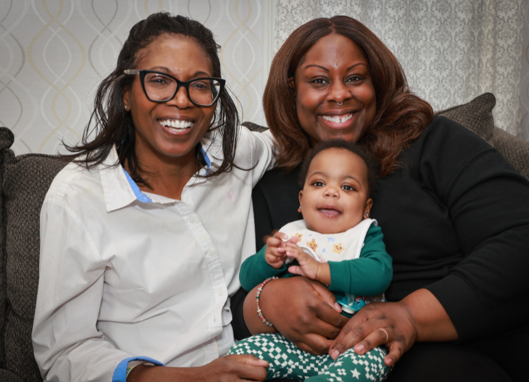
Explore stories of strength and inspiration from Valley Fertility Center patients.